
We recommend that all women over the age of 40 have regular breast exams and mammograms. The frequency of such examinations and the specific age at which screening should begin depend upon an individual’s risk factors for breast cancer, including personal and family medical histories.
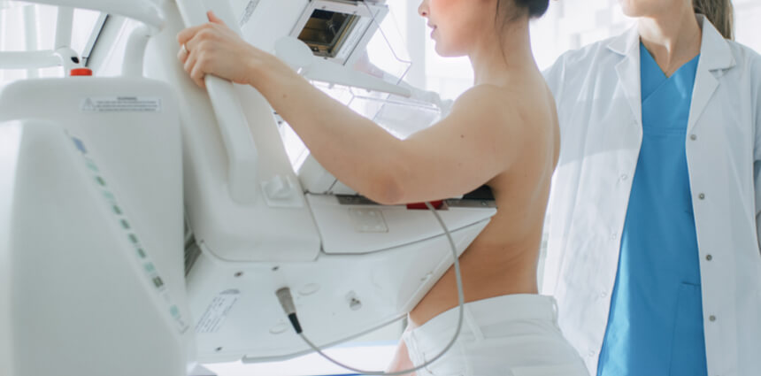
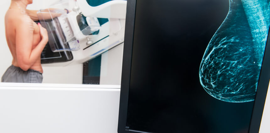
3D mammography, or breast tomosynthesis, is a relatively new breast imaging procedure, like traditional mammography, 3D mammography uses X-rays to produce images of breast tissue in order to detect lumps, tumors or other abnormalities. 3D mammography is capable of producing more detailed images of breast tissue.
3D captures multiple slices of the breast, all at different angles. The images are brought together to create crystal clear 3D reconstruction of the breast. The radiologist is then able to review reconstruction, one slide at a time, almost like turning pages in a book. This makes it easier for doctors to see if there’s anything to be concerned about.

Certain women at high risk for developing breast cancer may consider breast ultrasound in addition to screening mammography.
Ultrasound uses sound waves to produce images of internal organs and body tissue. Breast ultrasound is an imaging procedure that is not routinely used for screening, but it can be useful for evaluating some breast abnormalities identified by mammography. There is no radiation involved.
Breast ultrasound is primarily used to help physicians diagnose potential breast abnormalities previously detected by physical examination or mammography. Ultrasound can determine the difference between solid masses in breast tissue and fluid-filled cysts. It can also help determine whether a solid mass is a potentially cancerous tumor or a non-cancerous growth.
Many cancers are not visible on ultrasound and it is not intended to replace routine screening mammography. However, some physicians may recommend breast ultrasound in addition to screening mammography for certain women, such as those with dense breasts.
A woman receiving a breast ultrasound will be asked to remove clothing above her waist and she will be positioned on her back on an examination table. Ultrasound gel will be applied to the area of her breast being examined.
The radiology technologist performing the breast ultrasound will move a handheld device, called a transducer, over the examination area. The transducer tracks movements of sound waves through breast tissue and transmits this information to a computer, which renders it visually on a monitor in real time.
If a breast lesion has been detected, a breast biopsy may be required to determine its cause.
A breast biopsy is a medical procedure that extracts a tiny sample of breast tissue from the body for the purpose of testing. A breast biopsy may be performed in women who have detected a lump during physical examination, or in women whose screening mammography revealed a potential lesion. There are several different types of breast biopsy.
Breast imaging procedures help patients and their physicians detect potential abnormalities in breast tissue. Breast imaging procedures can also help physicians rule out cancer in some women who have detected a potential abnormality. However, a breast biopsy is required for physicians to make a conclusive breast cancer diagnosis.
Different types of breast biopsies are performed for different reasons. If a woman’s physician recommends a biopsy, she should ask why.
Breast biopsy experiences will vary based on the type of biopsy being performed. Image-guided biopsies use medical imaging technologies during the process to help physicians locate problem areas. The type of breast biopsy a physician recommends will depend on a patient’s specific circumstances, such as the location and size of a lesion in her breast.
In a fine needle aspiration breast biopsy procedure, an extremely thin needle attached to a syringe is used to extract a small amount of cells from a woman’s breast tissue.
Core needle biopsy uses a thin, hollow needle to extract tissue samples from the breast tissue. Prior to core needle biopsy, anesthesia will be injected into the target area. This numbs the breast of the woman being biopsied to prevent pain, but allows her to remain awake and alert during the procedure. Between two and six samples are extracted and each is approximately the size of a grain of rice.
Ultrasound may be used during fine needle aspiration biopsy or core needle biopsy to help a radiologist determine the exact location of the mass within a breast to be examined.
If you are a woman who needs a breast biopsy, or if you just want more information on your risk for breast cancer, contact us online and we will get back to you within one business day to schedule your appointment.